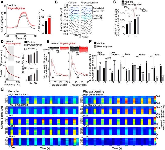
Figure 3.
Increased sensory-evoked CBF responses mirror increases in neuronal activity. A, Increased whisker-evoked-CBF responses by enhanced ACh tone (physostigmine) matched (B) increased of LFP evoked responses across cortical layers, delineated as the SL and GL layers and quantified by LFP (P1–N1) amplitude (C). Insets show averaged SL and GL evoked responses. D, Physostigmine altered the averaged baseline LFP power in SL (top) and GL (bottom), as quantified by the AUCs of the LFP spectra for high frequencies (20–60 Hz) and low frequencies (1–4 Hz), as indicated by gray boxes. E, Raw LFPs during stimulation (top) were increased by physostigmine in SL and GL. The normalized LFP spectra (bottom) further show shift of frequencies under enhanced ACh tone by physostigmine during sensory stimulation in both SL and GL. F, Power change during stimulation (as a percentage from baseline) in high-gamma (63–150 Hz) and low-gamma (30–57 Hz) bands showed a clear increase in all layers compared with control (vehicle). In contrast, the beta (15–30 Hz) band displayed no alteration of its power and power in lower-frequency bands [alpha (8–12 Hz) and theta (5–8 Hz)] decreased. G, Averaged band-limited power in selected frequency bands as a function of cortical depth for repeated trials (20 s baseline/prestimulation, 20 s 4 Hz whisker stimulation, 20 s poststimulation) in both control and physostigmine conditions. All data are shown as group averages. n = 78 and 67 trials for control and physostigmine conditions, respectively, from 8 rats. **p < 0.01; ***p < 0.001, Student's paired t test: physostigmine versus control (vehicle). a.u., Arbitrary unit; Stim, whisker stimulation.

