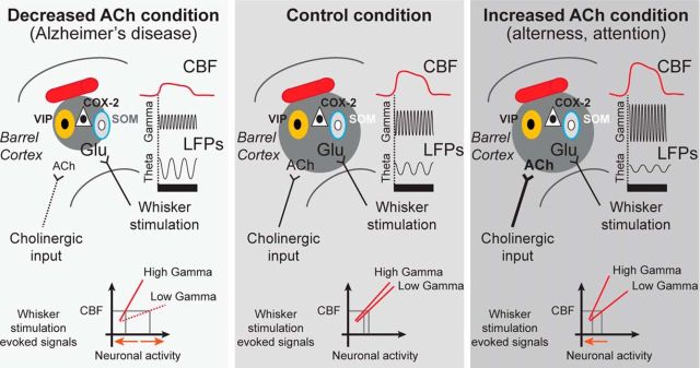
Figure 8.
Summary of whisker-evoked CBF and neuronal responses as a function of ACh tone. In the responsive control barrel cortex (dark gray circle), VIP interneurons (yellow cells) and COX-2-expressing pyramidal cells (white triangle) are typically activated (black nucleus, c-Fos positive), (middle panel) by the glutamate released upon whisker stimulation (black bar), whereas SOM interneurons (gray cells) remain largely inhibited (white nucleus, c-Fos negative). In conditions of increased ACh tone (as seen during arousal and attention conditions, right panel), the cortical neuronal circuitry is identical, but the resulting CBF and high-frequency oscillations are potentiated. In contrast, decreased ACh neurotransmission (mimicking AD, left panel) contracted the activated barrel cortex and altered the evoked CBF and neuronal responses. These changes involved lower amplitude of gamma bands and higher amplitude of low-frequency theta band, which results in altered fidelity of the correlations between LFP frequency bands and hemodynamics. Specifically, different levels of neuronal activity (orange arrows, bottom of panels) are needed for an equivalent whisker-evoked CBF response depending on ACh tone.

