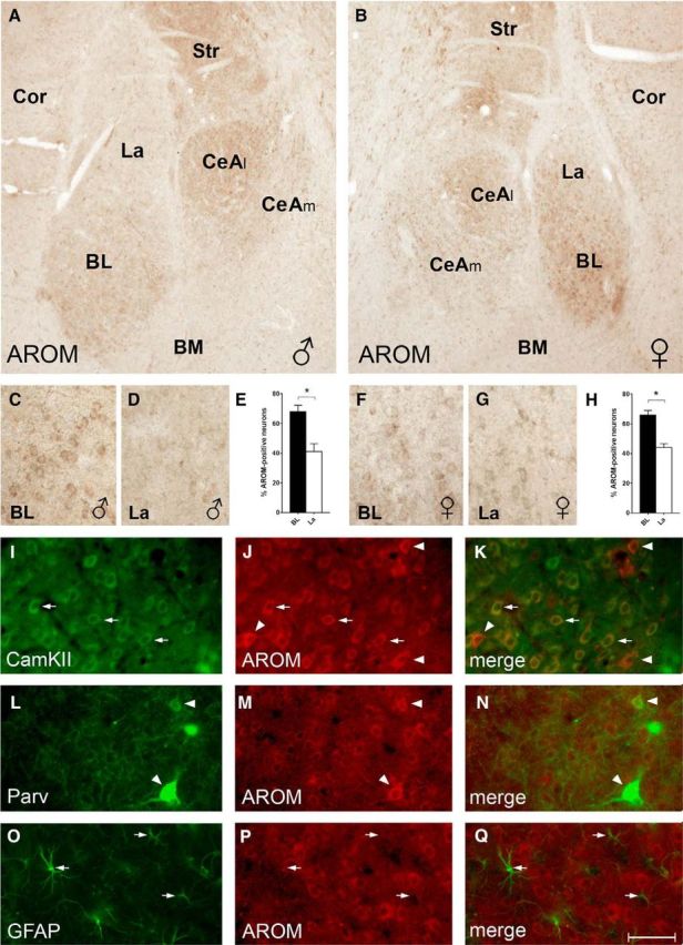
Figure 2.

AROM expression in rat BLA and CeA. A, B, AROM immunostaining of sections from a juvenile male (A) and female (B) rat reveals particularly robust AROM expression in subdivisions of the BLA and CeA: in the BLA, the BL had a higher density of AROM-expressing cells compared with the La and BM. In the CeA, the CeAl was more intensely labeled than the CeAm. C–H, Higher-magnification views of the sections shown in A (C, D) and B (F, G) demonstrate that within BLA the density of AROM-expressing cells is highest in BL, in which 68 ± 4% (E; males, n = 4) and 66 ± 3% (H; females, n = 4) of presumed neurons (nuclei > 5 μm) were immunoreactive for AROM. Density was significantly lower in La compared with BL both in males and females (41 ± 5% and 44 ± 3%, respectively; p = 0.02 for each sex). However, no significant differences were evident when densities between males and females were compared (p = 0.7 and p = 0.6, respectively, for BL and La). I–K, Coimmunostaining of AROM (red) and CaMKII (green) indicates that AROM expression is frequent in CaMKII-positive projection neurons in the BL (arrows). However, the presence of strongly AROM-immunoreactive neurons without CaMKII signal (arrowheads) suggests AROM expression also in interneurons. L–N, These interneurons include the Parv-expressing subpopulation, as Parv frequently colocalizes with AROM (arrowheads). O–Q, In contrast, no or only low levels of AROM signal were found in GFAP-immunopositive astroglial cells (arrows). Cor, Cortex; Str, striatum. Scale bar (in Q): A, B, 200 μm; C–G, 50 μm; I–Q, 25 μm.
