Figure 3.
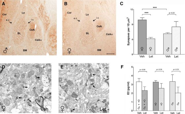
Reduced spine synapse density in BL in adult mice after letrozole treatment. A, B, Coronal sections through anterior amygdala of a young adult male (A) and female (B) mouse show that, as in the juvenile rats, AROM expression is more robust in the BL than in the La, whereas it is virtually absent in the BM. Similarly, the CeAl but not the CeAm of the central amygdala is intensely AROM immunoreactive. Arrows denote the external capsule (e.c.) and intermediate capsule (i.c.). C, Quantitative analysis of spine synapses after vehicle (Veh) or letrozole (Let) treatment, comparing males and females: letrozole caused a significant reduction of spine synapse density in the female mice (Veh, 9.2 ± 0.6 synapses/10 μm3; Let, 4.2 ± 0.4 synapses/10 μm3; p < 0.001; n = 6 each treatment), but not in the males (Veh, 5.6 ± 0.3 synapses/10 μm3; Let, 7.4 ± 1.4 synapses/10 μm3; p = 0.22; n = 6 each treatment). Notably, in the males, spine synapse density was already, under control conditions significantly lower than in the females (p < 0.001). D, E, Representative electron micrographs from the BL of a Veh-treated (D) and a Let-treated (E) female mouse. Note that spine synapses are present in both, but density is lower in the sections from the mouse treated with letrozole. F, Measurement of E2 plasma concentration after daily intraperitoneal injections of letrozole or vehicle in young adult male and female mice (n = 6, each sex and treatment) for 7 d suggest that letrozole reduces peripheral E2 concentration, but the difference did not reach significance (p = 0.09, n = 12 each treatment, if both sexes are considered; left bars). No difference in the response to letrozole was evident, when males and females were compared. (middle and right bars). Cor, Cortex. Scale bar (in B): A, B, 200 μm; D, E, 1 μm.

