Figure 8.

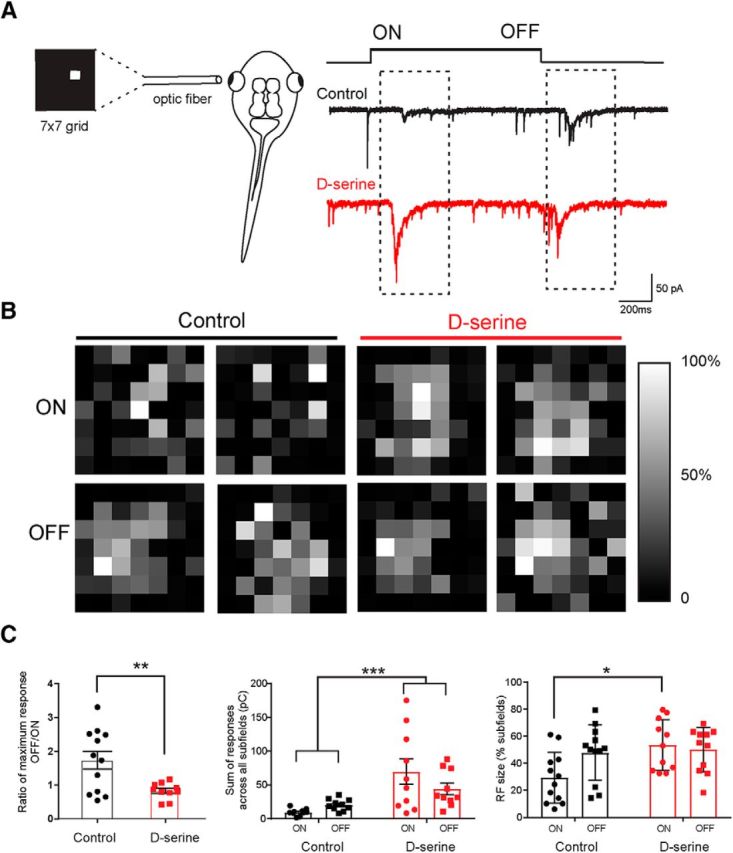
d-Serine increases receptive field size for ON responses. A, Schematic representation of the receptive field mapping setup. An optic fiber displaying a 7 × 7 grid of 49 individual subfields was aligned with the eye. Representative traces of whole-cell EPCSs recorded in the contralateral optic tectum in response to stimulus onset (ON) and stimulus offset (OFF) (control, black trace) (d-serine raised, red trace). B, Receptive field maps for tectal neurons in two example control cells and in two cells from animals raised in d-serine. Receptive fields were generated based on synaptic responses to bright ON stimulation in each subfield (top) and to the stimuli turning OFF (bottom) 1 s later. Receptive fields are plotted in grayscale. White represents the strongest subfield response. C, Left, Ratios of maximum CSC generated for an OFF response to the maximum CSC for an ON response show that the relatively stronger OFF response compared with the ON response consistently seen in control cells is equalized in d-serine cells (control, n = 12; d-serine, n = 10). **p < 0.001 (t test with one outlier removed by the ROUT outlier test). Middle, Sum of the CSCs evoked at all subfields is larger in animals raised in d-serine compared with control cells. ***p < 0.001 (two-way ANOVA, with Bonferroni post test; 3 cells removed by ROUT outlier test). Right, ON receptive fields are larger in animals raised in d-serine than in control animals. *p < 0.05 (two-way ANOVA with Bonferroni post test).
