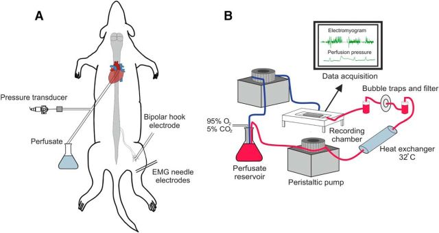
Figure 1.
Preparation setup. A, Schematic of animal setup with cannulation, recording, and stimulation sites shown. Animal is in the prone position. B, Components of the perfusion circuit and flow of ACSF. Red line represents flow from reservoir to preparation and the blue line represents the return flow. Two pumps are shown for clarity; all tubing actually inserts into a single pump.

