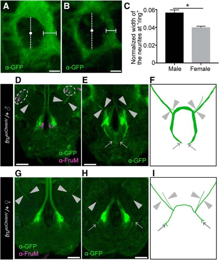
Figure 4.

Sexual dimorphisms in the fru-labeled circuitry in D. subobscura. A, B, Magnified images of the ring region boxed by a dotted white line in Figure 3E and F, respectively. The width of the medial portion of the inner fiber tract in the ring (indicated by horizontal bars) was measured along the lateral axis at the midpoint, which was defined as the midpoint (white dots) of the inner ring diameter along the dorsal-ventral axis (indicated by vertical broken lines). The width of the ring measured was normalized by the largest width of the dorsal central brain. Scale bars, 15 μm. C, A comparison of the normalized width of the ring fiber tract between the male (n = 5) and female (n = 3) frusoChrimV heterozygotes. The statistical significance of differences was evaluated by the Mann–Whitney U test: p = 0.0357. D–I, Sexual dimorphisms of the mAL neurites in the frusoChrimV heterozygous male and female. Scale bars, 30 μm. D, G, Magnified images of mAL cell bodies and neurites in the male (D) and female (G) brain. Cell bodies are encircled by a broken white line, and neurites are indicated by arrowheads. E, H, mAL neurites observed at a different plane, highlighting their tip portions (indicated by arrows) in the subesophageal ganglion (SOG) of the male (E) and female (H). The thick bilateral fiber tracts along the midline and large terminals in the SOG are of mcAL neurons. F, I, Schematic representation of the neurite projection pattern in the male (F) and female (I). Neurites are indicated by arrowheads. The dark oblique structure at the center represents the esophagus. Scale bars, 30 μm.
