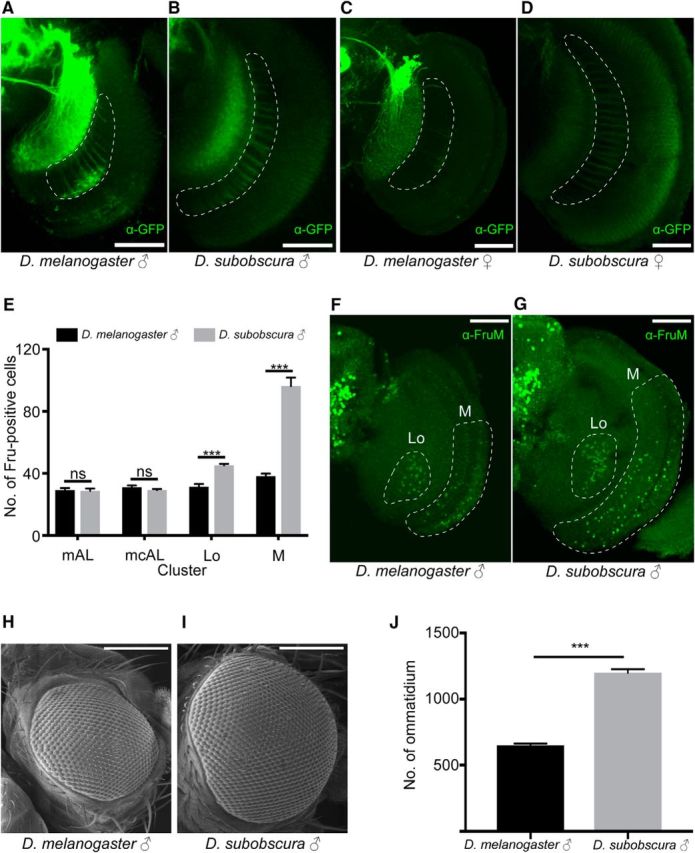
Figure 5.

Differences of the fru-labeled circuitry in the optic lobe between D. subobscura and D. melanogaster. A–D, The fru-labeled fibers of M neruons in the optic lobe (encircled with white broken lines) in D. melanogaster (A, C) and D. subobscura (B, D) in male (A, B) and (C, D). Scale bars, 50 μm. E, The number of anti-FruM antibody-immunoreactive cells in the mAL: p = 0.9213; mcAL: p = 0.6702; Lo: p = 0.0006; and M: p = 0.0006 clusters compared between D. melanogaster (n = 7) and D. subobscura (n = 7). The statistical significance of differences was evaluated by the Mann–Whitney's U test. ns, Not significant. ***p < 0.001. Error bars show SEM. F, G, Images of the optic lobe stained with the anti-FruM antibody in D. melanogaster (F) and D. subobscura (G) males. The region with Lo and M neuron somata is encircled with a white broken line. Scale bars, 50 μm. H, I, Scanning electron micrographs of the compound eye in D. melanogaster (H) and D. subobscura (I) males. J, The number of ommatidia composing a compound eye in D. melanogaster (n = 8) and D. subobscura (n = 8) males. The statistical significance of differences was evaluated by the Mann–Whitney's U test. p = 0.0006. Error bars show SEM.
