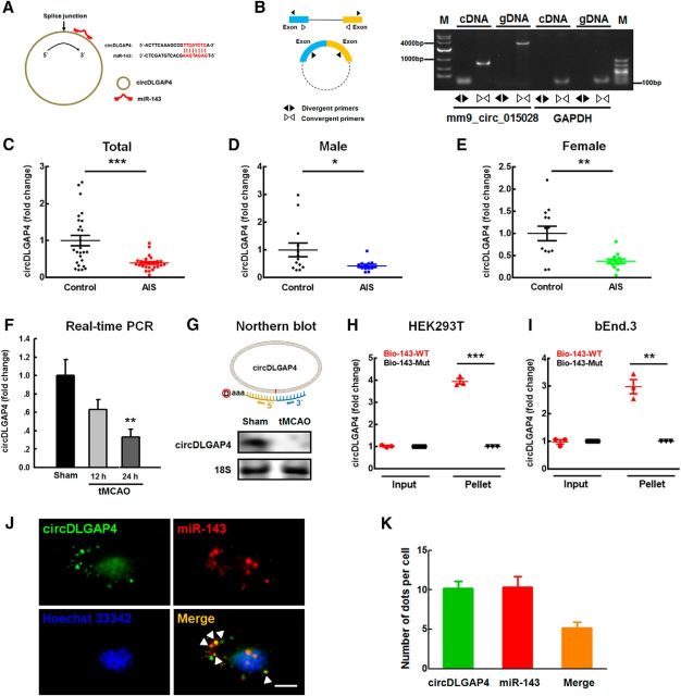
Figure 3.
circDLGAP4 binds miR-143 and is downregulated in the plasma of AIS patients and tMCAO mice. A, Left, circDLGAP4 contains one site that is complementary to miR-143 according to the bioinformatics program RNA hybrid. Right, A biotin-coupled miR-143 mutant. B, Divergent primers amplified circDLGAP4 from cDNA, but not genomic DNA (gDNA). GAPDH, linear control; M, marker. C, Examination of circDLGAP4 levels in the plasma of healthy controls and AIS patients via RT-PCR. circDLGAP4 levels were decreased in AIS patients by 0.6-fold the levels observed in healthy controls. n = 26 individuals/group. ***p < 0.001, AIS versus control (Mann–Whitney Test). D, E, circDLGAP4 levels in expression the plasma of male (D) and female (E) AIS patients were decreased compared with those in male and female healthy controls. n = 13 individuals/group. *p = 0.046, male AIS versus male control (Mann–Whitney Test). **p = 0.003, female AIS versus female control (Mann–Whitney Test). F, Effects of stroke on circDLGAP4 expression in the brain tissues of tMCAO mice. circDLGAP4 expression was determined at 12 and 24 h after tMCAO surgery via qRT-PCR. n = 8 mice/group. F(2,21) = 6.907, p = 0.102: 12 h tMCAO versus Sham. **p = 0.004, 24 h tMCAO versus Sham (one-way ANOVA followed by the Holm–Sidak Test). G, The expression of circDLGAP4 decreased in the mouse tMCAO mouse stroke model compared with that in sham group according to Northern blotting. H, I, circDLGAP4 was pulled down with biotinylated WT miR-143 (Bio-miR-143-WT) or mutant miR-143 (Bio-miR-143-mut). Biotinylated WT miR-143 (Bio-143-WT) or mutant miR-143 (Bio-143-mut) was transfected into HEK293T cells (H) and bEnd.3 cells (I). After streptavidin capture, circDLGAP4 and GAPDH mRNA levels were quantified via RT-PCR, and the relative immunoprecipitate/input ratios were plotted. Data are mean ± SEM of three independent experiments. HEK293T cells: ***p < 0.001, Bio-143-Mut Pellet versus Bio-143-WT Pellet. bEnd.3 cells: **p = 0.002, Bio-143-Mut Pellet versus Bio-143-WT Pellet (Student's t test). J, K, FISH hybridization of mature miR-143 and circDLGAP4 in brain endothelial cells (J) and quantitation of colocalization (K). White arrowheads indicate the colocalization of miR-143 and circDLGAP4. Green represents circDLGAP4. Red represents miR-143. Blue represents Hoechst 33342. Scale bar, 5 μm.

