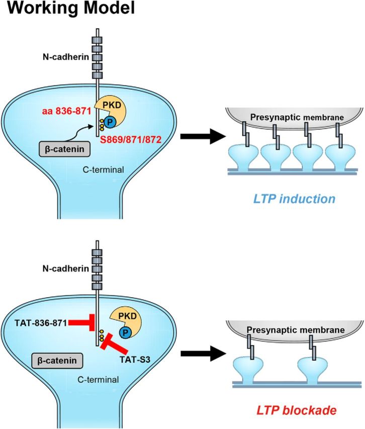
Figure 10.

Working model. PKD1 binds to N-cadherin at amino acid residues 836–871 and phosphorylates it at Ser 869, 871, and 872. Disruption of the modification of N-cadherin by PKD1 impairs the binding of N-cadherin to β-catenin and the membrane localization of N-cadherin, thereby inhibiting synapse formation and synaptic plasticity.
