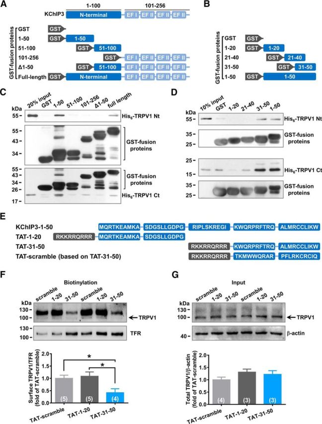
Figure 4.

KChIP3 N-terminal 31-50 fragment binds with both the N and C termini of TRPV1. A, B, Schematic diagram of the GST fusion proteins containing different fragments of KChIP3 (A) or KChIP3–1-50 (B). C, D, Representative results of GST-pull down assays. GST-1-50 precipitated both His6-TRPV1 N and C termini (C). More specificly, GST-31–50 precipitated both His6-TRPV1 N and C termini (D). E, Schematic diagram of TAT fusion peptides containing different fragments of KChIP3-1-50 and a scramble control of TAT-31-50. F, G, Representative results of biotinylation studies on the TRPV1 surface level in CHO-TRPV1 cells incubated with 3 μm TAT fusion peptides for 3 h. F, Bottom, Quantification analysis of the surface level of TRPV1. One-way ANOVA followed by Tukey's multiple-comparisons test, *p < 0.05. Total protein level of TRPV1 was also examined as well (G) and quantification analysis of the results is shown below (one-way ANOVA).
