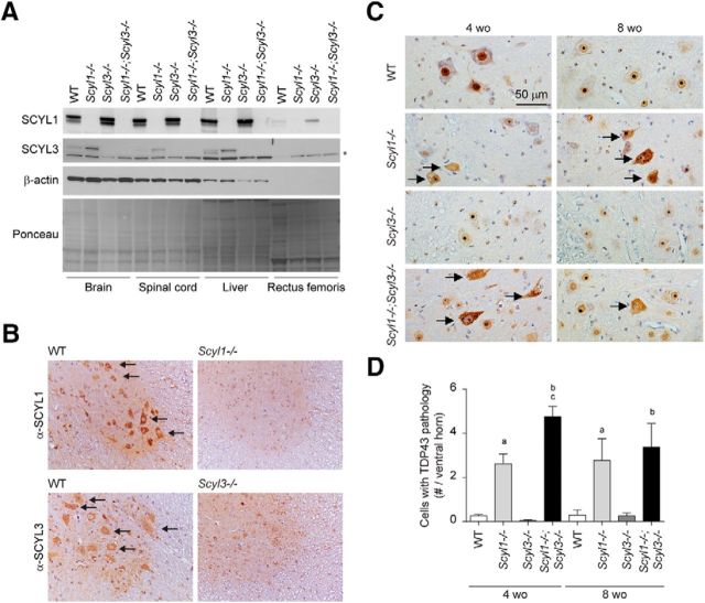
Figure 9.
Nuclear-to-cytoplasmic redistribution of TDP-43 in spinal motor neurons of Scyl1−/− and Scyl1−/−;Scyl3−/− mice. A, Expression of SCYL1 and SCYL3 in brain and spinal cord. Tissue distribution of SCYL proteins in WT, Scyl1−/−, Scyl3−/−, and Scyl1−/−;Scyl3−/− mice. Protein extracts prepared from various mouse tissues were resolved by SDS-PAGE and analyzed by Western blot, using antibodies against SCYL1, SCYL3, β-actin. Ponceau staining of the membrane is also shown as loading control. *Indicates nonspecific band. B, Overlapping expression of SCYL1 and SCYL1 in spinal motor neurons. Immunohistochemical staining of spinal cord cross-sections from WT, Scyl1−/− and Scyl3−/− mice, using antibodies against SCYL1 or SCYL3. Arrows indicate expression of SCYL1 or SCYL3 in spinal motor neurons. C, Immunohistochemistry using antibodies against TDP-43 on lumbar spinal ventral horn sections from 4- and 8-week-old WT, Scyl1−/−, Scyl3−/−, and Scyl1−/−;Scyl3−/− mice. Arrows indicate spinal motor neurons with relocalized TDP-43. D, Quantification of cells exhibiting TDP-43 pathology in ventral horn of 4- and 8-week-old WT, Scyl1−/−, Scyl3−/−, and Scyl1−/−;Scyl3−/− mice (n = 3 for each age group and genotype). Data are expressed as mean ± SEM. P values, determined by the one-tailed Student's t test, are indicated on the graph. a, WT versus Scyl1−/−, p < 0.05; b, WT versus Scyl1−/−; Scyl3−/−, p < 0.05; c, Scyl1−/− versus Scyl1−/−;Scyl3−/−, p < 0.05.

