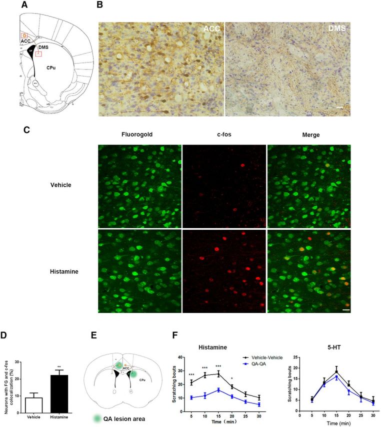
Figure 1.

Neuronal projections from ACC to DMS are involved in modulation of histamine-induced itch. A, Schematic depiction of neural projection from ACC to DMS. B, ACC neurons project to DMS revealed by tract tracing with BDA. Left, Tracer signals in ACC by BDA 3k retrograding from DMS. Right, Tracer signals in DMS by BDA 10k anterograding from ACC. The images were achieved from coronal brain sections around +0.62 mm anterior to bregma. Scale bar, 50 μm. C, D, c-fos immunostaining in FG-labeled ACC-DMS projections in response to histamine-induced itch. C, Representative immunostaining images of c-fos in FG-labeled ACC-DMS projections. FG was microinjected into DMS and the neurons in ACC was labeled by FG retrograding from DMS. D, Quantification of immunostaining images of c-fos in the total neurons labeled by FG. t(12) = 3.078, p = 0.0096. Student's t test. E, F, Disconnection of ACC and DMS projections with quinolinic acid attenuated histamine-induced, but not 5-HT-induced, itch. E, Schematic illustration of contralateral lesions of ACC and DMS. F, Scratching behavior induced by subcutaneous injection of histamine (500 μg/50 μl) rather than 5-HT (75 μg/50 μl) into the nape of neck was significantly decreased by disconnection of ACC-DMS circuit. Histamine: interaction, F(5,80) = 2.49;time,F(5,80) = 18.38. 5-HT: interaction, F(5,70) = 0.1175; time, F(5,70) = 13.29. Two-way ANOVA. All data are shown as mean ± SEM. *p < 0.05, **p < 0.01, ***p < 0.001 compared with vehicle or disconnection group. n = 8–10 mice for each group.
