Figure 5.
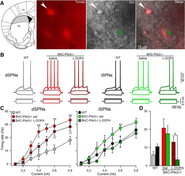
Intrinsic excitability is increased in both SPNs in BAC-Pitx3−/− mice. A, Scheme of a coronal slice depicting the striatal area without dopaminergic inputs in gray (left) and photographs (40×) of representative registered neurons in the gray area from a BAC-Pitx3−/− D1R-tomato mouse. B, Representative current-clamp recordings showing dSPNs and iSPNs recorded at 0.3 nA. C, Frequency of action potential evoked with depolarizing current in dSPNs and iSPNs. *p < 0.05; **p < 0.001 saline or l-DOPA BAC-Pitx3−/− mice vs WT mice; two-way ANOVA following Bonferroni's post-test. D, Histograms show the mean firing rate at 0.3nA to facilitate the comparison between dSPNs with iSPNs. *p < 0.05 dSPNs vs iSPNs t-test. Sal/sal, Saline; Str, striatum; Cx cerebral cortex.

