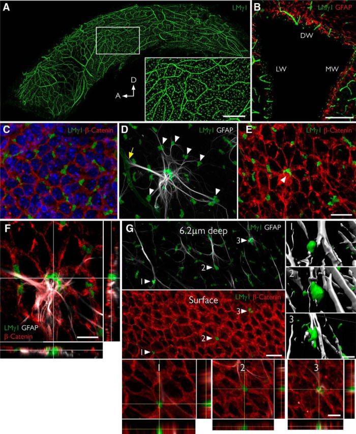
Figure 1.

Fractones are a major BM source in the SVZ and appear at the center of pinwheels. A, Whole mount of the lateral wall of the lateral ventricle of an adult mouse immunofluorescently stained for laminin γ1 (green). The frontal view reveals the profuse distribution of fractone bulbs. A higher magnification of the boxed area is depicted in the inset. B, Coronal section through the lateral ventricle, showing the uniform distribution of laminin γ1+ fractone bulbs (green) on all three walls: dorsal wall (DW), lateral wall (LW), and medial wall (MW). C, Frontal view of the ependymal cell layer, seen at 1 μm depth relative to the ventricular surface, stained for laminin γ1 (green) to visualize fractones and β-catenin (red) to visualize cell junctions in the ependymal layer. All fractone bulbs sit at the interface between cells. D, Maximum intensity projection of a Z-stack of a whole mount showing a GFAP+ NSC (gray) in contact with one blood vessel (yellow arrow) and >10 fractone bulbs (arrowheads) at the same time. E, The neural stem cell body also contacts a large bulb located at the center of a pinwheel (arrowhead). F, Orthogonal views revealing that the NSC–fractone interaction seen in C and D occurs at the surface of a pinwheel center. G, GFAP+ processes contact more bulbs at pinwheel centers (arrowheads) than bulbs at the basolateral surface of ependymal cells. Interactions between GFAP+ processes and fractone bulbs were labeled 1–3 and rendered, revealing that GFAP+ processes establish direct and complex contacts with fractones. Orthogonal views of these interactions confirm that fractone bulbs sit between ependymal cells at pinwheel centers and that GFAP+ processes directly contact them. Scale bars: A, 500 μm; A, inset, 200 μm; B, 200 μm; C–E, 25 μm; F, 12 μm; G, 25 μm; G, 1–3, 12 μm.
