Figure 2.

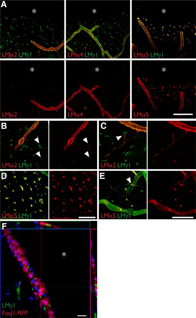
Fractone bulbs and fractone stems have distinct laminin compositions. A, Immunolabeling for laminin α2 (red, left), laminin α4 (red, middle), laminin α5 (red, right), and laminin γ1 (green) seen in maximum-intensity projections of thick (60 μm) coronal sections of young adult mice showing that fractone bulbs and blood vessels have distinct laminin compositions. Laminin α5 is the main α chain in fractone bulbs, followed by a faint presence of laminin α2. Laminin α4 is absent from fractones. B, Higher-magnification images showing the faint presence of laminin α2 in fractone bulbs (arrowheads). C, Higher-magnification images depicting fractone stems, rich in laminin α2 emerging from a blood vessel and contacting fractone bulbs (arrowhead). D, E, Higher-magnification images showing the ubiquitous presence of laminin α5 in fractone bulbs (D) and in its absence (E) in most of the fractone stems. F, Orthogonal views from a coronal section of a FoxJ1-RFP mouse showing that fractone bulbs are located at the ependymal layer only. Fractone bulbs may appear in thick stripes in coronal sections due the angle of the lateral wall and the thickness of the optical section. Asterisks indicate the ventricular cavity. Scale bars: A, 100 μm; B, D, 50 μm; C, E, 50 μm; F, 20 μm.
