Figure 4.
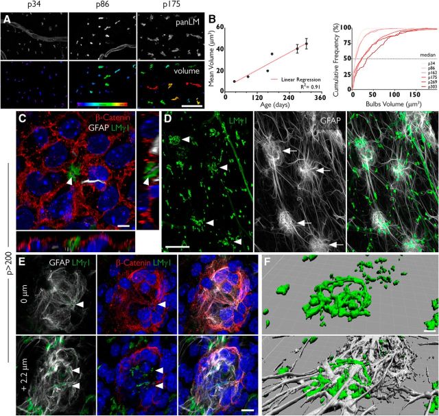
Fractones undergo major transformations with aging. A, Fractone bulbs, identified by immunofluorescent staining using a pan-laminin antibody (panLM; top), at three different ages. The volume of each individual bulb is color coded, showing that these structures increase in size with age. B, The mean volumes of fractones correlate with age (R2 = 0.91; left). An analysis of frequency showing that the median value for the volume of fractones also increases with age. C, A pinwheel in a whole mount of an 8-month-old mouse showing a tunneled fractone bulb at its center. Orthogonal views show a GFAP+ process (gray) sheathed by the fractone BM (green). D, Unusually large fractones (green, arrowheads) are associated with clusters of GFAP+ cells (gray, arrows). E, A detailed view of a cell cluster shows strong expression of β-catenin (red) in its margins and fractone fragments (arrowheads) in its interior. F, A tridimensional reconstruction of a giant fractone associated with a cell cluster showing how GFAP+ processes transverse these structures. Scale bars: A, 30 μm; C, 5 μm; D, 50 μm; E, 10 μm; F, 5 μm.

