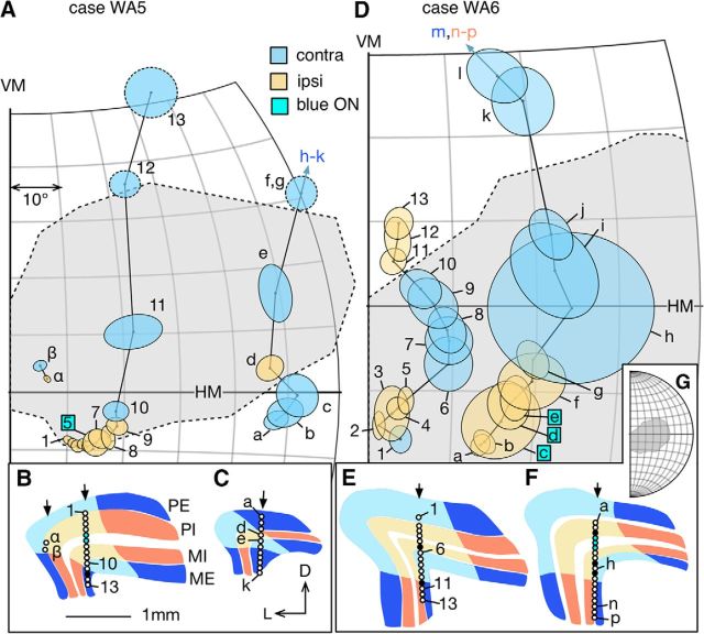
Figure 5.
A, D, Progression of receptive fields of LGN neurons sampled during vertical electrode penetrations in the adult group. The format is the same as in Figure 4. Locations of the recording sites are indicated in panels B, C (case WA5) and E, F (case WA6). In case WA5, the sections illustrated in B was 0.96 mm caudal to the one in C. In case WA6, the section illustrated in E was 0.48 mm caudal to the one in F. During the penetration illustrated in E, recording stopped at receptive field 13 without through the rest of the LGN. G, The relationship of the scotoma of WA6 to the visual hemifield.

