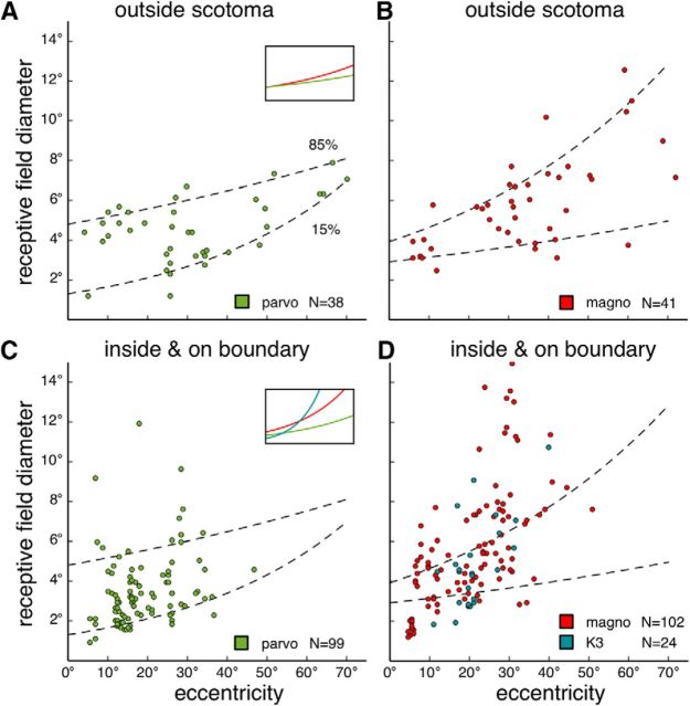
Figure 9.
Receptive field diameters as functions of eccentricity. The diameter was calculated as the diameter of a circular receptive field, whose surface area was matched to the surface area of the fitted oval-shaped receptive field (see Materials and Methods). Data pooled from all 6 cases. A, B, Receptive field diameters of units sampled in the putative parvocellular (A) and the magnocellular layers (B) outside the physiological scotomas. Dashed regression lines indicate values that account for 15% and 85% of the data. A, Inset, The 0.5 quantile regression lines. Green represents parvocellular units. Red represents magnocellular units. C, D, Receptive field diameters for units sampled in the parvocellular (C) and the magnocellular layers (D), inside or on the boundaries of the physiological scotomas. A small population of units sampled in the putative K3 layer is also plotted in D (as blue dots, in contrast to the red dots representing magnocellular units). Dashed lines are the same 0.15 quantile and the 0.85 quantile regression lines plotted in A and B. C, Inset, Regression lines for the median values. Green represents parvocellular units. Red represents magnocellular units. Blue represents K3 units.

