Figure 3.

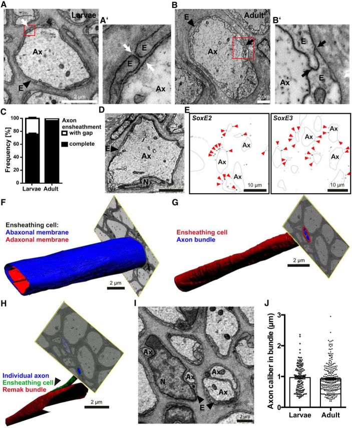
Axonal ensheathment by Schwann cell orthologs in the lamprey PNS. A, B, Electron micrographs of a cross-sectioned LLN in larval (A) and adult (B) lamprey. Note the glial ensheathment (E) of axons (Ax). Red boxes indicate areas magnified in A′ and B′, respectively. A′, B′, Axonal ensheathment occasionally displayed gaps (A′) in larval but was closed (B′) in adult lamprey. C, Quantification of gapped and closed axonal ensheathment in entire cross-sectioned LLNs. Note the maturation-dependent closure of ensheathment gaps. Data are shown as mean and SD. n = 787 axons in 2 larvae; n = 343 axons in 1 adult. D, Electron micrograph of an axon (Ax) in the cross-sectioned adult LLN highlighting an ensheathing cell nucleus (N). (E) FISH of a cross-sectioned LLN detecting SoxE2 and SoxE3, the lamprey orthologs of Sox9 and Sox10, respectively. Note that SoxE2 and SoxE3-labeling (pseudocolor representation by black puncta marked by arrowheads) partially outlines axons (Ax, black lines). F–H, FIB-SEM micrographs and 3D reconstruction. F, Adaxonal (red) and abaxonal (blue) plasma membrane of a representative cell ensheathing an individual axon in the larval LLN. Note the structural homogeneity over at least 20 μm. See also Movie 1. G, Bundle of multiple axons (blue) ensheathed by a single ensheathing cell (red) showing homogeneity over at least 20 μm. See also Movie 2. H, As an anecdotal observation, an axon (blue) with its individual ensheathment (green) leaves a bundle of multiple axons (red). I, Electron micrograph of a LLN highlighting bundles of two or more axons (Ax). For quantification of axons per bundle, see Figure 4. E, Ensheathing cell; N, nucleus. J, Calibers of LLN axons ensheathed in bundles. Data are shown as mean and SEM. n = 120 axons in 2 larvae; n = 274 axons in 1 adult.
