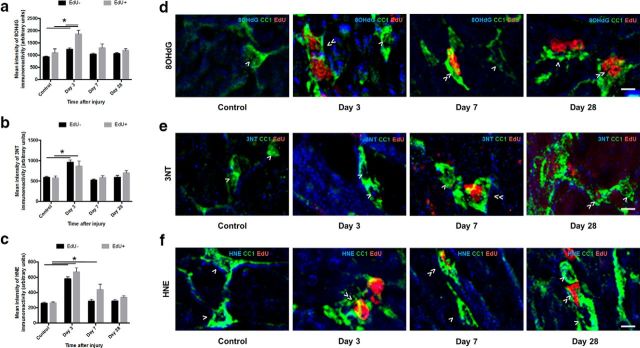
Figure 7.
8OHdG-, 3NT-, and HNE-derived fluorescence in mature oligodendrocytes vulnerable to secondary degeneration in vivo. Mean ± SEM fluorescence intensity within mature oligodendrocytes (CC1+ cells) is shown following 8OHdG (a), 3NT (b), and HNE (c) labeling in uninjured optic nerve and at 3, 7, and 28 d following partial transection. Arbitrary values for immunofluorescence were categorized as EdU− (black bars) or EdU+ (gray bars) to discriminate oligodendrocytes present at the time of injury and oligodendrocytes derived after injury. Representative images of 8OHdG (d), 3NT (e), and HNE (f) labeling are of mature oligodendrocytes located within the ventral optic nerve vulnerable to secondary degeneration. d–f, Cells indicated are CC1+/EdU− (>) or CC1+/EdU+ (≫). Statistical comparisons were made across and between groups. *p ≤ 0.05, differences in immunointensity compared with controls. Scale bar, d–f, 10 μm.

