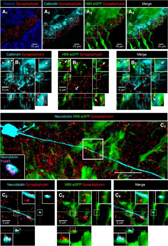
Figure 3.

V1R make synaptic-like contacts with motoneurons (HB9-eGFP) at E12.5. A, Coronal slice of the lumbar spinal cord of E12.5 HB9-eGFP mouse embryo with cell nucleus staining (HOESCH) and synaptophysin immunostaining (A1). A1, Note that synaptophysin immunostaining is mainly restricted in the ventral funiculus. A2, Calbindin staining showing the distribution of V1R neurite extensions and synaptophysin immunostaining. A3, eGFP immunostaining showing the distribution of MN neurite extensions and synaptophysin immunostaining. Antibody against GFP was used to visualize MN morphology better (Czarnecki et al., 2014). A4, Superimposition of eGFP fluorescence, calbindin immunostaining and synaptophysin immunostaining (A1–A4 are confocal stacks). B, Single confocal sections with z projections of enlarged images from A2 to A4 showing the colocalization of synaptophysin punctates with calbindin immunostaining opposed to eGFP immunostaining (B1–B3; enlarged images in boxes). B2, Note that a synaptophysin punctate colocalized with calbindin immunostaining (B1, arrow) did not colocalize with eGFP immunostaining (B2, arrow). Note that synaptophysin punctate colocalized with eGFP immunostaining (B1, arrowheads) did not colocalize with calbindin immunostaining (B2, arrowhead). Barred arrow (B1) shows a colocalization of calbindin and synaptophysin immunostaining not opposed to eGFP immunostaining. B4, superimposed images (B1–B3) with z projections showing calbindin immunostaining and eGFP appositions. C1, Confocal stacks showing neurobiotin-injected Foxd3-immunoreactive V1R, HB9-eGFP immunostaining and synaptophysin immunostaining in an SC open book preparation. C2–C4, Single confocal sections with z projections of enlarged images from C1 (white box) showing the colocalization of synaptophysin punctates with neurobiotin staining (C2), the apposition of the same synaptophysin punctates to eGFP (C3) and the apposition of neurobiotin staining containing synaptophysin punctates to eGFP (C4; enlarged image in boxes in C2–C4), indicating the presence of a V1R synaptic-like contact on an MN neurite.
