
Figure 4.
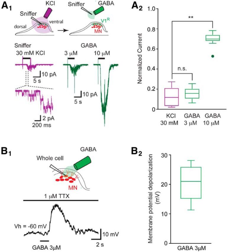
Paracrine release of GABA detected by a sniffer outside-out patch. A1, Top drawing shows the location of the outside-out sniffer to detect paracrine release of GABA (left) and to obtain outside-out currents in response to GABA application (left). Bottom traces (purple): example of outside-out sniffer current evoked by the application of 30 mm KCl when the sniffer electrode was positioned in the dorsal area of the SC close to motor columns. Enlarged trace shows single-channel currents at the onset of the sniffer current. Right traces (green) show outside-out current evoked by the application of 3 and 10 μm GABA to a sniffer patch positioned outside the spinal cord. Purple and green traces are from the same outside-out patch. A2, Box plots of normalized maximum outside-out current evoked by KCl application (purple left) and by the application of 3 or 10 μm GABA (green right) on the same outside-out sniffer patch (n = 9). Amplitudes of the outside-out currents evoked by the application of 30 mm KCl, 3 or 10 μm GABA, were normalized to the amplitude of the outside-out currents evoked by the application of 30 μm GABA (data not shown). Note that the normalized amplitude of the outside-out current evoked by 30 mm KCl application (0.118 ± 0.097) was not significantly different (p > 0.9) from the normalized amplitude of the outside-out current evoked by the application of 3 μm GABA (0.152 ± 0.062). Normalized amplitudes of the outside-out currents evoked by the application of 30 mm KCl or of 3 μm GABA were significantly different (KCl: p = 0.0029; 3 μm GABA: p = 0.00665) from the normalized amplitude of the outside-out currents evoked by the application of 10 μm GABA (0.697 ± 0.073). **p < 0.01. B1, Example of motoneuron membrane potential depolarization evoked by the application of 3 μm GABA in the presence of 1 μm TTX (current-clamp recording: Vh = −60 mV; ECl = −30 mV). B2, The application of 3 μm GABA evoked a depolarizing response of 20.6 ± 6.1 mV (n = 7).