
Figure 5.
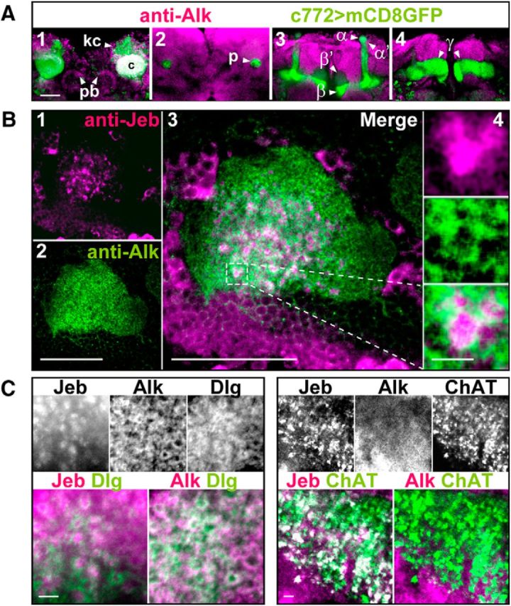
dAlk and its ligand Jeb are enriched in synapses of projection neurons and Kenyon cells. A, dAlk is enriched in MB calyces but is absent in the MB lobes. Confocal images of single optical sections of the fly brain from posterior to anterior (A1–A4) were acquired at the level of MB calyces (A1), pedunculus (A2), α/β lobes (A3), and γ lobe (A4). Confocal images were acquired at the same section levels with identical settings. dAlk was visualized with an anti-dAlk antibody (purple). Fly brain structures were marked with a membrane GFP (green) encoded by the mCD8:GFP transgene expressed with the neuronal c772-GAL4 driver. White represents colocalization of dAlk and GFP immunofluorescence. dAlk protein accumulates within the dendrites (A1: calyces, c) and cell bodies (A1: Kenyon cells, kc, arrowhead) of MB neurons and in the protocerebral bridge (A1: pb, arrowheads). It is also expressed widely in the neuropil, above background. In contrast, MB axons (A2: pedunculus, p, arrowhead) (A3: α, α′, β, and β′ lobes, arrowheads) (A4: γ lobes, arrowheads) are devoid of dAlk staining. Scale bar, 50 μm. B, dAlk and Jeb display a complementary pattern of expression within MB calyces. Confocal images of a single optical transverse section of an MB calyx were acquired using identical settings and at the same section level corresponding approximately to its middle section (B1–B3). Jeb protein was visualized with an anti-Jeb antibody (B1, purple), and dAlk protein was visualized with an anti-dAlk antibody (B2, green). Scale bar, 50 μm. Colocalization of Jeb and dAlk immunofluorescence (B3, merge, white) is shown at the same magnification. Scale bar, 50 μm. Inset, Higher magnification of the hatched box, showing in a single synaptic microglomerulus (B4), from top to bottom, Jeb, dAlk, and their complementary pattern. Scale bar, 10 μm. C, dAlk is expressed at the postsynaptic dendritic active zones of the MB calyces, whereas Jeb is expressed presynaptically in the synaptic buttons of apparent projection neurons. Confocal images of single optical sections of MB calyces were acquired at similar levels, using identical settings and displaying triple staining of Jeb, dAlk, and membrane GFP. Scale bars, 10 μm. Left, White represents signals of anti-Jeb, anti-dAlk, and anti-Dlg, a marker of the postsynaptic active zones. Colored images represent lack of colocalization of Jeb (purple) and Dlg (green), and colocalization (white) of dAlk (purple) and Dlg (green). Right, White represents signals of anti-Jeb, anti-dAlk, and anti-ChAT, a marker of presynaptic terminal buttons. Colored images represent complete colocalization of Jeb (purple) and ChAT (green), and lack of colocalization (white) between dAlk (purple) and ChAT (green).