Figure 12.

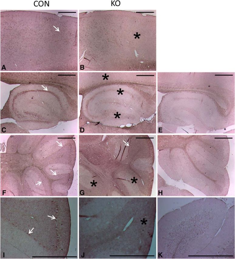
In situ hybridization histochemistry of glut3 gene expression in adult mouse brain. After overnight incubation with DIG-labeled riboprobes targeting exon 6 of the glut3 gene, alkaline phosphatase-conjugated sheep anti-DIG IgG antibody (Roche Applied Science) was used for color development. In CON (A, C, F, I), antisense probes show positive signals (arrows) in the neuronal cells of cortex, hippocampus, and cerebellum. In KO (B, D, G, J), antisense probes show significantly reduced immunoreactivity including within the parietal cortical association and retrosplenial agranular cortex (B), CA1 and dentate gyrus region of hippocampus (D), and cerebellar lobule (G, J). Controls with the corresponding sense RNA probes show the immunoreactivity completely abolished (E, H, K). Asterisks indicate reduced immunoreactivity in B, D, G, and J. Scale bars, 500 μm.
