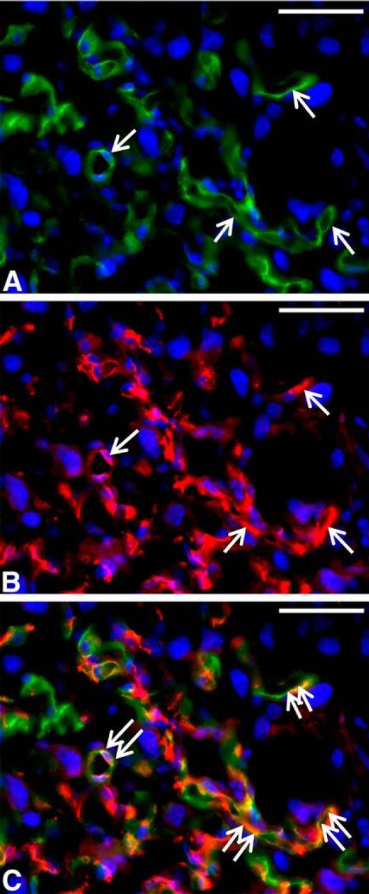
Figure 5.

Immunofluorescence localization of CD34 and Nestin proteins in E18 placenta. Alexa Fluor 488-conjugated donkey anti-rabbit IgG (1/250) and Alexa Fluor 594-conjugated donkey anti-goat IgG (1/250) were applied after the rabbit anti-CD34 (1/50) and goat anti-Nestin antibodies (1/50, each) over a 3 h incubation at room temperature. DAPI served as the nuclear stain. CD34 (A) and Nestin (B) proteins were expressed in the membrane of endothelial cells (arrows). A double-exposure image shows colocalization of CD34 (green) and Nestin (red) in endothelial cells of fetal capillaries (double arrows in C). Scale bars, 50 μm.
