Figure 9.
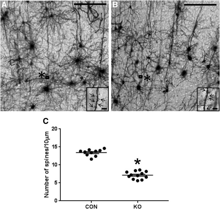
The number of apical dendritic spines was counted in the five continuous segments, each 10 μm in length, starting at 50 μm distance from the boundary between the dendrite and soma of pyramidal neurons in the cortex at PN15. A magnified view from the rectangle (*) is shown in the right corner. Arrows indicate spines. The number of spines was significantly reduced in the apical dendrites of KO mice (B) compared with CON (A). C shows number of spines per 10 μm, n = 12 neurons, n = 4 animals (KO), n = 10 neurons, n = 4 animals (CON) (unpaired t test, t statistic [df: 20] = 14.99, *p < 0.0001 vs CON). Scale bars, 200 μm each. Magnified view demonstrates dendritic spines (arrows). Scale bars, 5 μm each.

