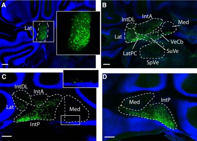
Figure 1.
Sox14 marks a subset cells in cerebellar nuclei. P21 Sox14Gfp/+ coronal sections show the cerebellum from rostral to caudal. A, Only the lateral cerebellar nucleus is seen in rostral sections. Sox14+ cells are found in the cerebellar nucleus, and these cells are distributed unevenly, seen clearly in the magnified image (inset). B, The lateral nucleus merges with the interposed nuclei and the vestibular nuclei: SuVe, superior vestibular nuclei; VeCb, vestibulocerebellar nuclei. There are Sox14+ cells throughout, except in the dorsal parts of the medial nucleus. C, More caudally, the lateral and anterior interposed nuclei recede so to only occupy a small dorsolateral domain, whereas the posterior interposed nucleus takes over. Small numbers of Sox14+ cells are seen in the ventral edge of the medial nucleus (inset). D, Most caudally, the medial nucleus is seen clearly as an almond shape above the posterior interposed nucleus. Though the shape of the nucleus is well defined by the background staining, again no Sox14+ cells are seen in this region. Lat, Lateral nucleus; LatPC, parvicellular lateral nucleus; IntDL, dorsolateral interposed nucleus; IntA, anterior interposed nucleus; IntP, posterior interposed nucleus; Med, medial nucleus; VN, vestibular nucleus; RN, reticular nucleus. Scale bar, 200 μm.

