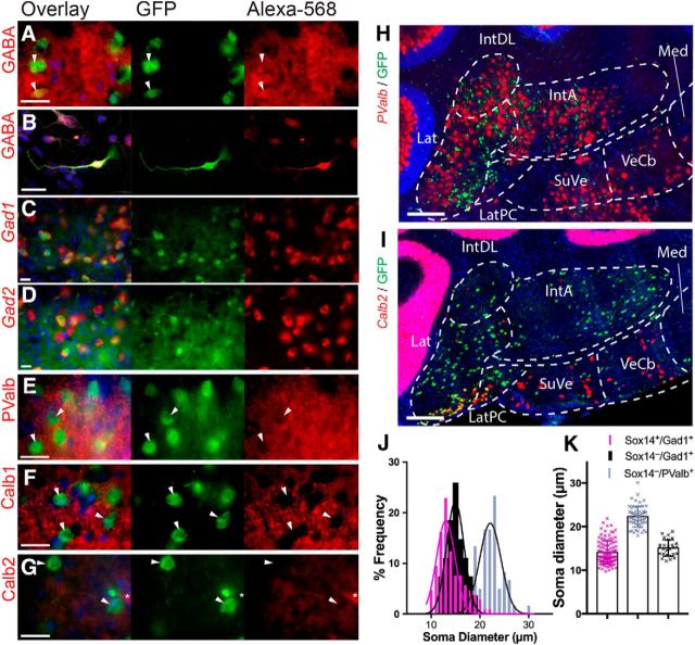
Figure 2.
In the cerebellar nuclei, Sox14 are small, exclusively GABAergic, PV-ve neurons GFP versus PValb, GANA CALB1,2, GABA, GAD1, and GAD2. A–I, Comparison of Sox14:GFP with other known cell markers by immunohistochemistry or in situ hybridization in Sox14Gfp/+ P21 sections (A, C–I) and primary cell culture of brain tissue from Sox14Gfp/+ P0 neonates (B). Immunostaining for GABA (A, B), MAP2 blue label in (B), Gad1 (C), Gad2 (D), PValb (E), Calb1 (F), and Calb2 (G), imaged at 100× (40× for in situ hybridization) magnification of the lateral nucleus. The columns show the overlay, then GFP only and Alexa Fluor 568 only. The white arrowheads show examples of GFP+ cells that colocalize with GABA, Gad1, and Gad2 but not PValb, Calb1, or Calb2. There is little immunoreactivity for Calb2 within the cerebellar nuclei (G), but a single GFP−/Calb2+ cell is seen, denoted with an asterisk. H, I, In situ hybridization against PValb (H) and Calb2 (I) demonstrates the distribution of the different cell types. Lat, Lateral nucleus; LatPC, parvicellular lateral nucleus; IntDL, dorsolateral interposed nucleus; IntA, anterior interposed nucleus; Med, medial nucleus; SuVe, superior vestibular nuclei; VeCb, vestibulocerebellar nuclei. H, PValb expression is observed in complementary large nuclear cells to GFP+ cells. I, Calb2 expression is mostly observed in two distinct populations within the Sox14+ cells of the cerebellar nuclei. Sox14−/Calb2+ are seen in the central parts of the lateral nucleus, whereas Sox14+/Calb2+ exist in a dense cluster in the ventral parts of the lateral nucleus. J, K, Scatterplot (J) and histogram (K) of soma size as measured by mean soma diameter (in micrometers). The mean soma diameters are as follows: GFP+/Gad1, 14.1 ± 0.3 μm; GFP−/Gad1+, 15.1 ± 0.4 μm; and GFP−/PValb+, 22.3 ± 0.3 μm (mean ± SEM). The peak frequency for cell diameter of both GFP+ and GFP− Gad1 populations is very similar. In addition, the larger GFP+ cells overlap with the PValb+ population, demonstrating that size is not a sufficient determinant of cell type.

