Figure 5.
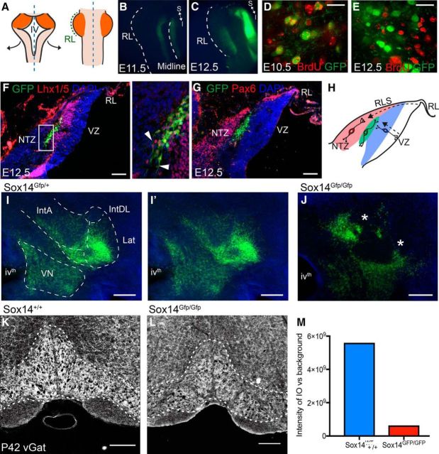
Integration of inhibitory projection neurons into the nuclei is mediated by Sox14. A, Sox14Gfp/+ hindbrains were opened up dorsally along the midline and mounted flat so that the rhombic lip, which originally lined the intersection between the cerebellum and roof plate, is the most lateral edge (in green); the cerebellar anlage is in orange. B, C, GFP expression is seen at E11.5 (B) and E12.5 (C) on either side of the midline, whereas expression in the cerebellar plate is only seen from E12.5 onward. D, E, BrdU birth-dating analysis. Scale bars, 20 μm. GFP+ cells colocalize with BrdU that was injected at E10.5 (D), whereas BrdU injected at E12.5 (E) shows no colocalization, indicating that all the GFP+ cells are born before E12.5. F, In situ hybridization against Lhx1/5 and GFP in the Sox14Gfp/+ E12.5 sagittal brain sections. The Lhx1/5-expressing cells span the anteroposterior axis of the cortical transitory zone and are mostly Purkinje cell precursors. However, there is a dorsal layer of the Lhx1/5+ and GFP+ population that are genetically distinct, as shown in the higher-magnification image (inset). These cells appear to be in a tangential orientation (white arrowheads), unlike the GFP−/Lhx1/5+ Purkinje cells that are migrating radially from the ventricular zone. G, Pax6+ cells migrating along the rhombic lip migratory stream toward the nuclear transitory zone sit dorsal to the GFP+ cells. H, A schematic to show the tangential orientation of the GFP+ cells (green) alongside the Pax6 excitatory cells migrating tangentially along the subpial rhombic lip migratory stream (RLS, red) and the GFP−/Lhx1/5+ Purkinje cells that are migrating radially from the ventricular zone. I, J, Coronal sections of the Sox14+ cells in the developing CN of P0 Sox14Gfp/+ mouse (I) compared with the Sox14Gfp/Gfp knock-out mouse (J). Scale bars, 200 μm. I′, The same image without borders drawn to highlight that for the Sox14Gfp/+ mouse, the migratory streams already resemble the future boundaries between the subnuclei, whereas for the knock-out, the cells fail to populate some areas, leaving large gaps (asterisks) and deviant clusters of cells. There are still some likenesses between the two brains, which suggest that there are other migratory mechanisms at work in the development of nucleo-olivary neurons. K–M, Density of vGAT labeling in the inferior olive of an adult wild-type (K) and Sox14 mutant (L) mouse shows a difference in signal to background intensity (M). Scale bars: F, 100 μm; I–L, 200 μm. RL, Rhombic lip; Lat, lateral nucleus; IntDL, dorsolateral interposed nucleus; IntA, anterior interposed nucleus; VN, vestibular nucleus; VZ, ventricular zone; ivth, fourth ventricle.

