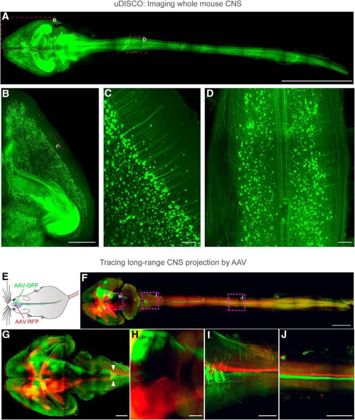
Figure 2.

uDISCO enables investigation of long-range neuronal connections spanning the entire mouse body and entire CNS in unprecedented detail (Pan et al., 2016). A–D, The entire CNS (whole brain and spinal cord) of a Thy1-GFP-M mouse was cleared by uDISCO and imaged by LSFM. Because of the superior preservation of the fluorescent protein signals achieved by uDISCO, detailed neuronal structures are clearly visible. Zoom-in view of the brain hemisphere and spinal cord within boxed region in A are shown in B and D, respectively. Zoom-in view of B is shown in C, where individual pyramidal cell bodies and dendrites are clearly resolved. Scale bars: A, 10 mm; B, 1 mm; C, 100 μm; D, 1 mm. E–J, AAV2-Syn-GFP was transduced in the right motor cortex of mice, and AAV2-Syn-RFP in the left motor cortex. The brain was then cleared by uDISCO and imaged with LSFM. Fluorescence expression of the virally delivered proteins was detectable in the brain and throughout the intact spinal cord (F–J). Details of neuronal extensions of boxed regions in F are shown in H (thalamus and midbrain), I (cervical), and J (thoracic spinal cord regions). Arrowheads in G indicate decussation of the descending motor axons. Scale bars: F, 5 mm; G, 2 mm; H–J, 1 mm.
