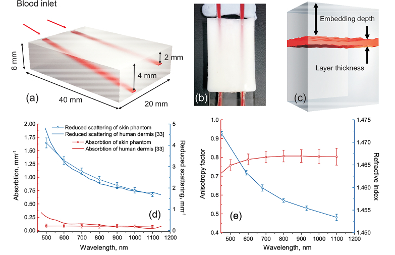
Fig. 2.
3D schematics of the biotissue phantom with the embedded blood vessels (a); photograph (top view) of the manufactured phantom with the channels filled with fully oxygenated blood (b); three-layer tissue phantom model used for calculations of diffuse reflectance spectra (c); measured optical properties of the biotissue phantom (d,e).

