
Figure 2.
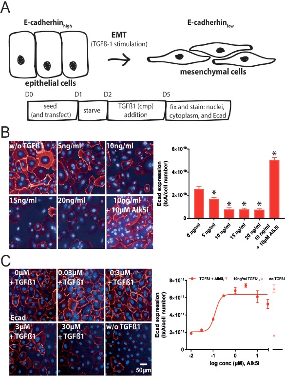
Phenotypic EMT assay. A. Schematic representation of the EMT assay with time points of treatment and readouts. At the end of the assay cells were prepared for high content imaging on E-cadherin expression. B. Images of hSAEC treated with increasing doses of TGFβ-1 and stained for nuclei and Ecad. Images were acquired with an Opera Phenix High Content Screening System (right). Ecad/ total cell numbers was quantified with a custom protocol using Columbus software and plotted against cumulating doses of TGFβ-1. From this data 10 ng/ml TGFβ-1 for all further experiments was chosen. C. Immunofluorescence images of hSAECs stimulated with 10 ng/ml TGFβ-1 and progressive concentrations of the Alk5i SB-525334 (right). The Alk5i dose-dependently inhibited the TGFβ-1-mediated reduction of Ecad expression an IC50 of 0.12 μM (left). *P < 0.05 versus non-TGFβ-1 treated cells.