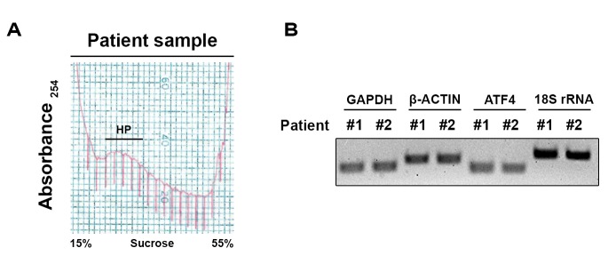
Figure 2.
Representative polyribosome profile obtained from prostate transurethral resection and RT-PCR of polyribosomal RNA. A. Prostate tissue samples were collected and homogenized. The homogenates were then sedimented on a 50% sucrose cushion. The polyribosome-enriched resultant pellet was resuspended and loaded on a 15%–55% sucrose gradient and the polyribosome profile was obtained by continuous UV monitoring at 254 nm during fractionation of the gradient. RNA was extracted from pooled heavy polyribosomes (HP), and its quality and integrity was validated to be suitable for RNA-Seq as above. B. RNA isolated from HP of cancerous (#1) and benign (#2) prostate specimens was then analyzed by qRT-PCR using oligos specific to GAPDH, β-ACTIN, ATF4 and 18S rRNA. The amplified PCR products were then migrated on an agarose gel.

