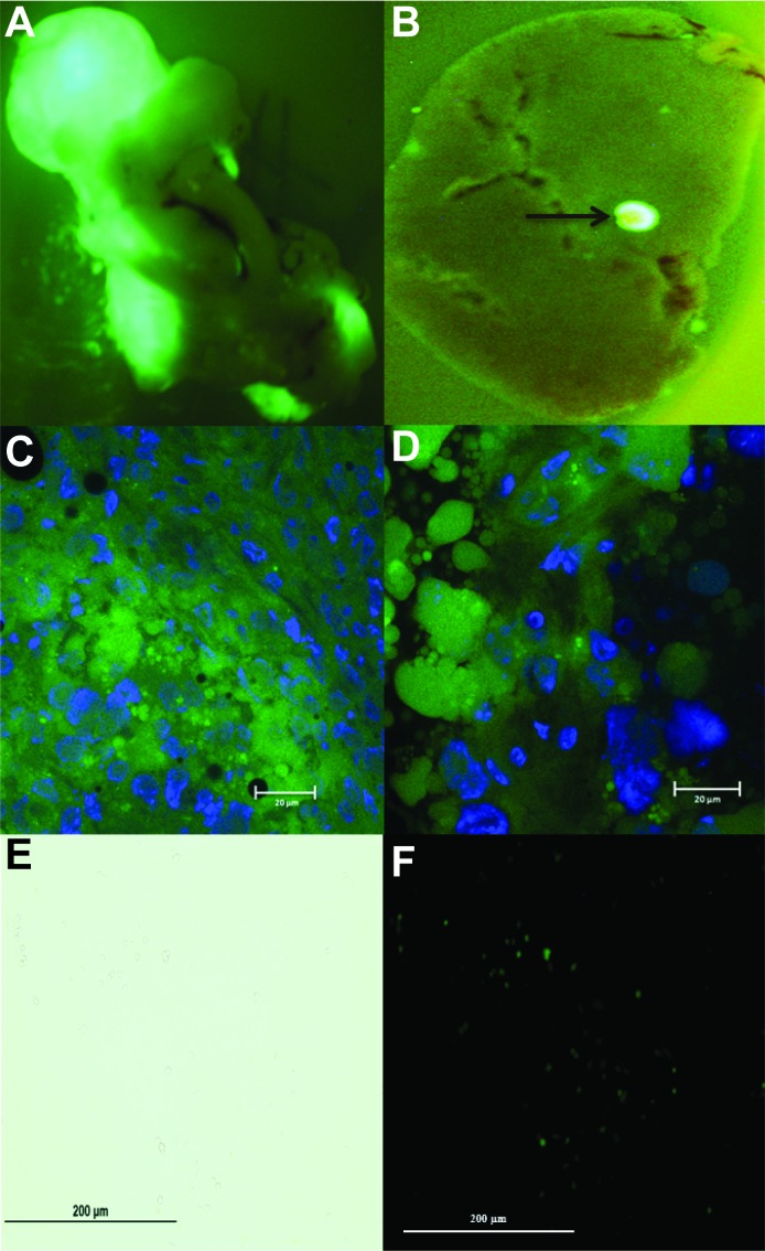
Figure 3. Ex vivo pancreatic and liver GFP fluorescent imaging for primary and metastatic tumor identification.
A. Immediately following cardiac draw and mouse sacrifice, the pancreas and liver were removed for ex vivo imaging under an Olympus Fluorescent Stereoscope to confirm primary pancreatic tumor from GFP expressing MIA PaCa-2 cells. B. Liver metastasis from GFP expressing MIA PaCa-2 cells. Arrow corresponds to liver metastasis. C. GFP immunofluorescent staining of primary pancreatic tumor. D. GFP immunofluorescent staining of liver metastasis. Specimens were fixed in OCT for immunofluorescent staining, cut into 10 µm section using a MICROM HM 505E, mounted in VectaShield Mounting Medium with DAPI and viewed with a Zeiss 710 Laser Scanning Confocal Microscope. Green areas represent positive GFP staining within the cell cytoplasm in both pancreatic and liver specimens. Blue areas represent cell nuclei. E. Bright field. F. GFP expression. Liver specimens with visible metastatic lesions were used to isolate single tumor cells to test for the presence of GFP expression. Hepatic sections were quickly minced with a scalpel in cold HBSS and centrifuged before the addition of 0.5% w/v collagenase. Cells were plated and allowed to attach before being examined for GFP expression at 20× magnification.

