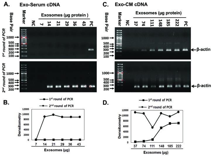
Figure 2. Amplification of exosomal β-actin by two-step PCR.
Agarose gels were ran after first and second round of PCR amplification as shown in upper and lower panel respectively. A. Exo-serum cDNA (ALL patient serum) failed to amplify sufficient β-actin product after first round of PCR. B. PCR band intensity of (Fig. 2A) measured by image J densitometry. C. Exo-CM cDNA (JM1 cell line) did show a detectable amount of β-actin amplicon after first round of PCR. D. PCR band intensity of (Fig. 2C) measured by image J densitometry. Both Exo-serum cDNA and Exo-CM cDNA β-actin amplicons were detected after second round of PCR. A minimal exosomal amount of 14–37 µg was sufficient to obtain a signal only after two rounds of PCR. NC: negative control, no template added; PC: positive control, cell-cDNA used as template.

