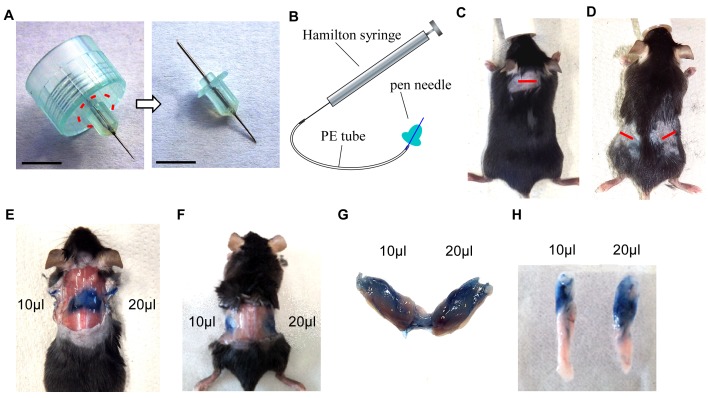
Figure 1.
Set up of injection device and establishment of protocol. A. Original pen needle (dashed line indicates the cutting area) (left) and needle after cutting off plastic rim (right). Scale bar = 0.5 cm. B. Illustration showing setup of injection device. C and D. Pictures of anesthetized and shaved mouse, incision sites are marked by a red line in the interscapular region (C) or at the flanks, proximal of hips (D). E. Situs after injection of 10 μl and 20 μl Trypan blue solution in BAT. F. Situs after injection of 10 μl and 20 μl Trypan blue solution in the upper region of WATi. G. Dissected BAT from (E). H. Dissected WATi from (F).

