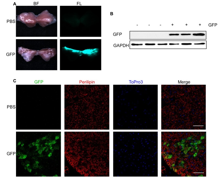
Figure 2.
GFP fluorescence in interscapular BAT after lentivirus injection. A-C. 1000 ng RT of lentivirus carrying GFP under control of a CMV promoter (GFP) in 25 μl HBSS or 25 μl PBS were injected into each interscapular BAT lobe of 4-week-old male mice and analyzed after 1 week. A. Bright field (BF, left) and fluorescent images (FL, right) of PBS (upper panel) and GFP (lower panel) injected BAT. B. GFP expression assessed by Western blotting. Respective blots of GFP and the loading control GAPDH are shown. C. GFP expression assessed by immunohistological staining. PFA-fixed BAT sections were triple stained with antibodies directed against GFP (green) and Perilipin (red) as well as with ToPro3 (blue) to stain the nuclei. Scale bar = 50 µm.

