. 2019 Jul 30;8:e46500. doi: 10.7554/eLife.46500

Figure 1. Architecture of the HNH domain ββα-Me fold in different binding forms of Cas9 (a–d) and site-directed mutagenesis experiments identifying potential catalytic residues (e–f).

(a–d) ββα-Me fold in the sgRNA-bound state of Cas9 (a), in the intermediate state (b), in the pre-catalytic state (c), and in the pseudoactive state (d). The ββα-Me fold is represented as pink ribbons, and the residues are shown in stick models and colored by atom type (C, dark green; N, blue; O, red). If present, the bound Mg2+ ion is depicted as a magenta sphere, and only the tDNA phosphate-sugar backbone is displayed for clarity. The location of the Cas9 D861 is highlighted by an arrow, and the dashed lines denote hydrogen bonds or coordinative bonds. (e) The expression and DNA-editing activity of the wild-type and D861A variants of Cas9 paired with an sgRNA sequence that targets the egfp gene in HEK293T-EGFP cells. (f) The expression and DNA-editing activity of the wild-type and indicated variants of Cas9 paired with an sgRNA sequence that targets the egfp gene in HEK293T-EGFP cells. The retention of EGFP expression reflected the loss of activity of Cas9 protein in the cells.

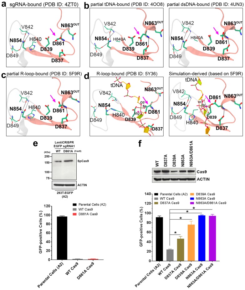
Figure 1—source data 1. Numeric data for activity quantification of Cas9 and its variants (XLS).
elife-46500-fig1-data1.xlsx (284.6KB, xlsx)
DOI: 10.7554/eLife.46500.009

Figure 1.

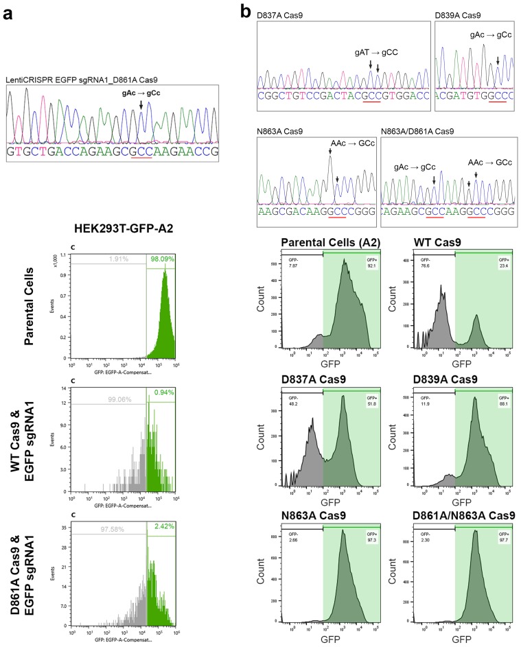
Figure 1—figure supplement 1. The DNA sequencing analysis of vectors for expressing different Cas9 variants and the flow cytometry analysis of HEK293T-EGFP cells with different Cas9-sgRNA expression vectors.

Figure 1—figure supplement 1.

(a) Upper panel: A DNA sequencing chromatogram showing the D861A mutation of the Cas9 gene open reading frame in the lentiCRISPR expression vector of Cas9 with the EGFP sgRNA1 sequence. Lower panel: Representative histograms of flow cytometry analysis in HEK293T-EGFP cells with the expression vectors of wild-type (WT) and D861A Cas9. (b) Upper panel: DNA sequencing chromatograms showing the D837A, D839A, N863A and D861A/N863A mutations of the Cas9 gene open reading frames in the lentiCRISPR expression vectors of Cas9 with the EGFP sgRNA1 sequence. Lower panel: Representative histograms of flow cytometry analysis in HEK293T-EGFP cells with the expression vectors of WT, D837A, D839A, N863A and D861A/N863A Cas9.

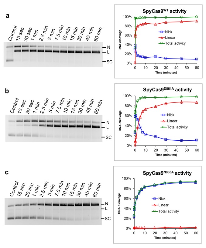
Figure 1—figure supplement 2. Plasmid cleavage activity of SpyCas9WT.

Figure 1—figure supplement 2.

(a) SpyCas9D861A (b) and SpyCas9N863A (c) Left panels: The gel images of DNA electrophoresis showing the bands of different plasmid conformations due to the cleavage activity of Cas9. Right panels: The quantitative results of cleaved DNA over the indicated reaction time. Each data point presents the average value of three replications. Error bars represents standard error of mean. The results indicate that SpyCas9D861A has a similar activity profile as SpyCas9WT, expect for a slower rate. SpyCas9N863A produces only nicked products even after one hour of reaction. [N: nicked, L: linear, SC: supercoiled].

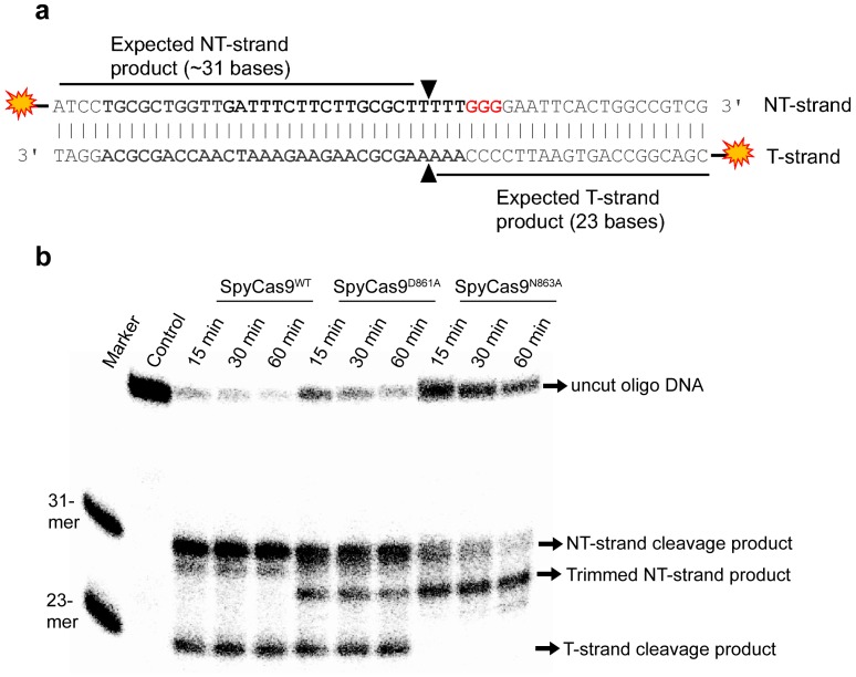
Figure 1—figure supplement 3. Cleavage of radioisotope-labeled oligo DNA substrate by SpyCas9WT, SpyCas9D861A and SpyCas9N863A.

Figure 1—figure supplement 3.

(a) A schematic illustration of radioisotope-labeled oligo DNA substrate used in this study. The duplex is labeled with 32P at the 5’-end of both strands. The sequences in bold are the protospacer; the PAM sequence is in red. Black triangles indicate the anticipated cleavage sites by HNH and RuvC. The size of denatured DNA products as visible in electrophoresis followed by film exposure is indicated for each DNA strand. (b) A representation of gel image showing the products of oligo DNA cleavage. Reactions were performed for different time periods, and the products were resolved on a denaturing 16% urea-formamide gel. The NT-strand cleavage product and T-strand cleavage product are produced respectively by the RuvC and HNH domains. SpyCas9N863A lacks T-strand cleavage product, suggesting that the HNH activity is eliminated in this variant. In summary, our in vitro cleavage reactions of plasmid and oligo DNAs clearly indicate that N863 is indispensable for the HNH nuclease activity, whereas D861 appears to provide a supporting role to enable a faster reaction rate but is nonessential for target DNA strand cleavage. A close inspection of our simulation trajectories led us to propose that D861 might have a role in stabilizing the catalytic ββα-Me motif by forming an intra-molecular salt bridge (Figure 2d), or aid initial recruitment of metal ions around the active center with other negative species like D839 and D837 (Figure 2a).

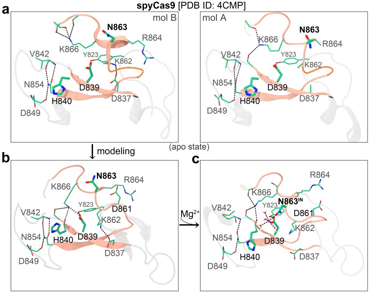
Figure 1—figure supplement 4. The architecture of the HNH domain ββα-Me motif in the apo structure of SpyCas9 (a) and structural modeling of SpyCas9 with Mg2+ ion bound at the catalytic center (b–c).

Figure 1—figure supplement 4.

The ββα fold is depicted as a pink ribbon diagram, and the residues at and around the fold core are shown as a stick model and colored by atom types (C, dark green; N, blue; O, oxygen). The disordered loop in apo-SpyCas9 is indicated by a chain of beads. The magenta sphere represents the bound Mg2+, and the dashed lines denote hydrogen bonds or coordination bonds.

Figure 1—figure supplement 5. The architecture of the HNH domain ββα-Me motif in the apo structure of Actinomyces naeslundii Cas9 (AnaCas9) (a) and in that of partial dsDNA-bound Staphylococcus aureus Cas9 (SauCas9) (b).

Figure 1—figure supplement 5.

The ββα fold is depicted as a pink ribbon diagram, and the residues at and around the fold core are shown as a stick model and colored by atom types (C, dark green; N, blue; O, oxygen). The magenta sphere represents the bound Mg2+, and the dashed lines denote hydrogen bonds or coordination bonds.