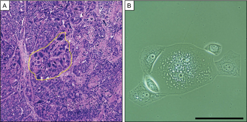
Figure 3. Polyploid giant cancer cells in prostate cancer in vivo and in vitro.
(A) H&E image of a lymph node prostate cancer metastasis with PGCCs (one region indicated by yellow border). (B) Phase image of a PC3 PGCC undergoing asymmetric division to form mononuclear and typical-sized daughter cells. PC3 cells were cultured with 10nM Docetaxel for 3 days followed by 4 days in Docetaxel-free media. (scale = 200 um)

