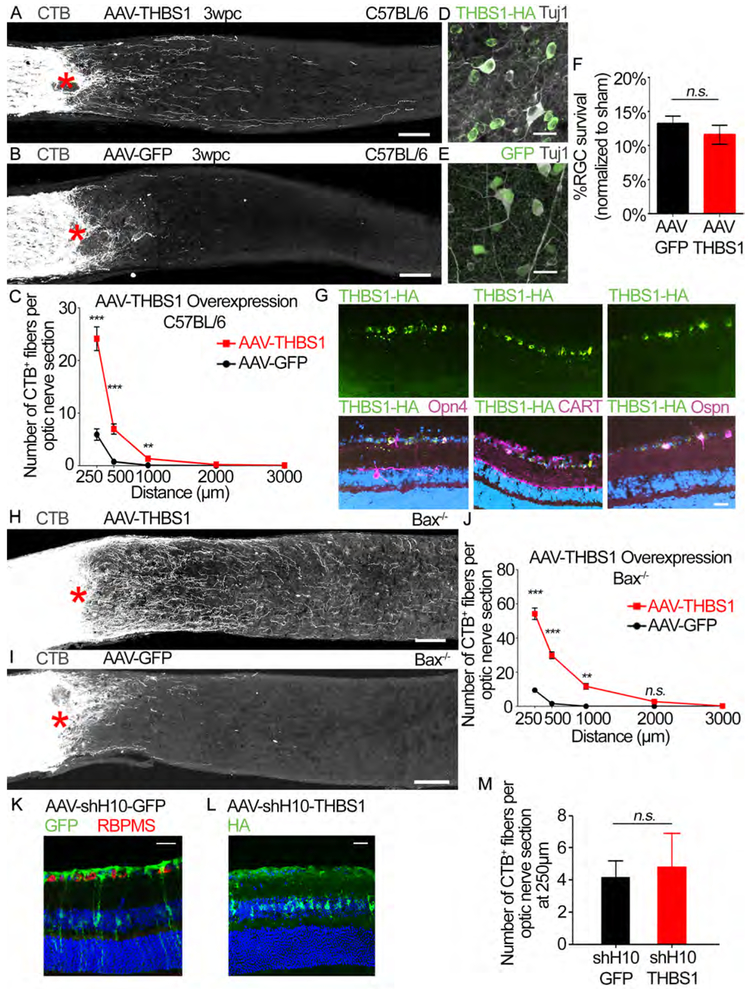
Figure 5: Ectopic THBS1 expression promotes RGC axon regeneration.
(A and B) Images of optic nerve sections showing CTB-labeled axons (grey) in C57BL/6J mice injected with either (A) AAV-THBS1 or (B) AAV-GFP. Asterisks, lesion site. Scale bars, 100 μm.
(C) Quantification of axon regeneration for (A and B). Average number of CTB+ fibers per optic nerve section at indicated distances from the lesion. n=10 per condition
(D and E) Representative retina whole-mount images showing Tuj1+ (grey) and (D) THBS1-HA (green) or (E) GFP labeled cells in C57BL/6J mice shown in (A and B). Scale bars, 25 μm.
(F) Quantification of RGC survival for (D-E). Average % survival of RGCs (Tuj1+ RGCs) in injured retina normalized to uninjured (sham) retina. n=10 per condition.
(G) Representative retinal sections from AAV-THBS1-HA injected mouse stained with antibodies against HA (green) and indicated RGC subtype markers (magenta; Opn4, CART and Ospn which are markers of ipRGCs, DSGCs and alpha RGCs, respectively). Dapi in blue. Scale bar, 25 μm.
(H and I) Images of optic nerve sections showing CTB-labeled axons (grey) in Bax−/− mice injected with either (H) AAV-THBS1 or (I) AAV-GFP. Asterisks, lesion site. Scale bars, 100 μm.
(J) Quantification of axon regeneration for (H and I). Average number of CTB+ fibers per optic nerve section at difference distances from lesion site. n=5 per condition.
(K) Representative retinal section from AAV-shHIO-GFP injected mouse stained with GFP (green) and RBPMS (RNA-Binding Protein With Multiple Splicing, a RGC marker; red) antibodies. Dapi in blue. Note that GFP expression is predominantly in non-RGCs. Scale bar, 25 μm.
(L) Representative retinal section from AAV-shH10-THBS1-HA injected mouse stained with HA antibody. Dapi in blue. Scale bar, 25 μm.
(M) Quantification of axon regeneration for the AAV-shH10 animals in (K and L). Average number of CTB+ fibers per optic nerve section at 250 μm from the lesion site. n=6 for AAV-shH10-GFP and 7 for AAV-shH10-THBS1.
Statistics, (C, and J) Unpaired t-test, 2 tailed, at each distance; (F and M) Unpaired t-test, 2 tailed, n.s. p>0.05, *p≤0.05, **p≤0.01, ***p≤0.001, Error bars, SEM.

