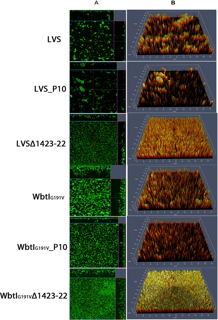
Figure 2.
Comparison of biofilm formation by F. tularensis LVS, WbtIG191V (LPS O-Ag mutant), LVSΔ1423-22 (CLC mutant), and O-Ag and CLC double mutant before and after passage in CDMB by confocal laser scanning microscopy. Panel (A) contains orthogonal sections showing horizontal (z) and side views (x and y) of reconstructed three-dimensional biofilm images at a magnification of 25x. Biofilms were stained with Syto9 showing both live and dead bacterial cells. Panel (B) shows topographical images of biofilm growth of the parent LVS and its surface structure mutant strains. Loss of surface carbohydrate was associated with denser biofilms, but subculture passage in CDMB resulted in thinner biofilms.

