Figure 5.
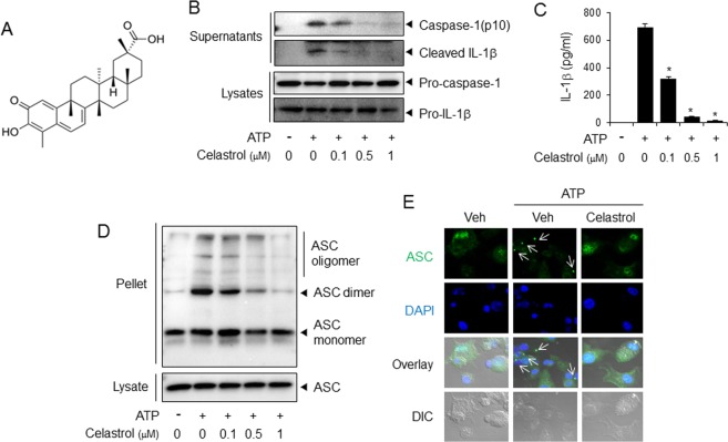
Celastrol inhibits ATP-induced activation of NLRP3 inflammasome in primary macrophages. (A) Chemical structure of celastrol. (B–E) Primary mouse macrophages were primed with LPS (100 ng/ml) for 4 hr. The cells were treated with celastrol for 1 hr and then stimulated with ATP (5 mM) for (B,D,E) 1 hr or (C) 2 hr. (B) The cell culture supernatants and cell lysates were immunoblotted for pro-caspase-1, caspase-1(p10), pro-IL-1β, and IL-1β. (C) The cell culture supernatants were analyzed for secreted IL-1β using ELISA. The values represent the means ± SEM (n = 3). *Significantly different from ATP alone, p < 0.05. (D) Cell lysates were processed for immunoblotting of ASC. (E) The cells were stained for ASC (green). The nuclei were stained with 4′,6-diamidino-2-phenylindole (DAPI; blue). The arrows indicate ASC speckles. The data are representative of three independent experiments. For immunoblotting results, the cropped blots from full length gels were presented. DIC, differential interference contrast.

