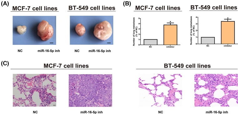
Figure 3. MiR-16-5p promotes breast cancer growth in vivo.
(A) The tumor growth of mice in each group is detected after MCF-7 and BT-549 cell lines are subcutaneously inoculated (bar = 2 mm). (B) Metastatic nodes in lungs in different groups are counted after MCF-7 and BT-549 cell lines are injected through tail veins. (C) H&E staining images of lung tissues after MCF-7 and BT-549 cell lines are injected through tail veins (original magnification, ×200). The data are indicated as mean ± SD. *P≤0.05, Student’s t test.

