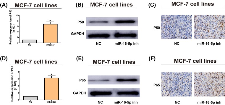
Figure 5. Down-regulation of miR-16-5p promotes the pathway of NF-κB pathway.
(A) MiR-16-5p inhibitors are transfected into MCF-7 cells and the mRNA level of P50 is assessed by qRT-PCR. (B) P50 protein level after MCF-7 cells are treated with miR-16-5p inhibitors are examined via Western blotting, with GAPDH as control. (C) P50 protein level in nude mice with tumor after MCF-7 cells treated with miR-16-5p inhibitors are inoculated that are detected via IHC. (D) MiR-16-5p inhibitors are transfected into MCF-7 cells and the mRNA level of P65 is assessed via qRT-PCR. (E) P65 protein level after MCF-7 cells are treated with miR-16-5p inhibitors is analyzed via Western blotting, with GAPDH as control. (F) P65 protein level in nude mice with tumor after MCF-7 cells treated with miR-16-5p inhibitors are inoculated is examined via immunohistochemistry (IHC). The data are indicated as mean ± SD. *P≤0.05, Student’s t test.

