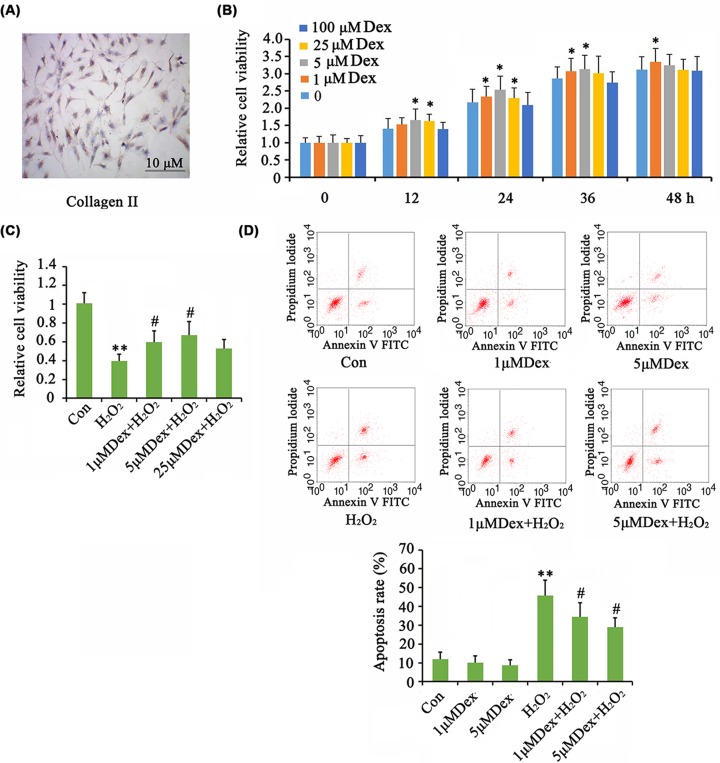
Figure 1. Dex improved the proliferation of AF chondrocytes in the presence or absence of H2O2.
The isolated AF chondrocytes were identified by ICC assay (A). Chondrocytes were treated with doses of Dex for different time periods to determine the optimum concentration and culture time. Then cell viability was tested (B). Oxidative stress was induced by treating the cells with 1 mM H2O2 for 1 h. Afterward, cells were cultured in fresh medium or the medium with the supplementation of Dex with further incubation for 24 h. Then cell viability (C) and apoptosis rate (D) were tested. *P<0.05 and **P<0.01 vs. control group; #P<0.05 vs. the group that was treated with H2O2 alone.

