
FIG. 3.
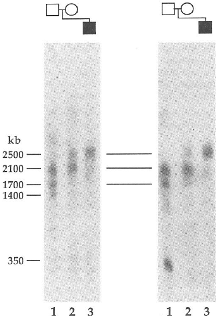
PFGE provides evidence for the lack of a submicroscopic deletion in PWS patients with uniparental disomy. NotI fragments are shown for probes IR4-3R (D15S11) (left) and 34 (right) in PWS2 (lane 3), M2 (lane 2) and F2 (lane 1). Multiple bands are indicative of partial cleavage at restriction sites when CpG is methylated32 and a similar pattern of multiple bands has been observed in other normal controls (results not shown). PWS2 shares a normal 2,500-kb band with his mother, although in the latter this results from partial digestion at a NotI site in this region of the genome. The same 2,500-kb band has been detected in other normal individuals. The larger three fragments are common to probes IR4-3R, 189-1 (results not shown) and 34, showing that these loci are closely linked in region 15q11q13. These probes and a 1,300-kb fragment detected with probes 3-21 and IR10-1 (unpublished data) are deleted in all PWS10 and AS6 patients with cytological deletions. Additional PFGE analysis (results not shown) also indicates that PWS2 displays PFGE restriction fragments of normal size for IR10-1 in a NotI digest (1,300 kb) and for IR39d in a BssHII digest (250 kb). PWS1 also seems to be intact for the probe 34 and 3-21 loci because the restriction fragments detected using NotI and BssHII are unaltered compared with those of normal individuals.
METHODS. PFGE analyses were performed as described previously33 using a modification of techniques for DNA preparation and digestion in agarose blocks34. Electrophoresis was performed in 0.8% agarose in TBE (89 mM Tris buffer, pH 8.0, 89 mM boric acid, 2 mM EDTA) at 22 °C and was divided into three 48-h intervals during which the forward polarity pulse durations were varied from 75-600 s, 600-2,500 s and 2,500-3,000 s and field intensities were set at +2, +1.7 and +1.3 V cm−1, respectively. The ratios of the duration and intensity of the inverse polarity pulse relative to the forward were 0.4 and 0.5, respectively.