
Figure 1.
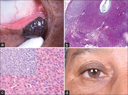
(a) Clinical photograph of the right eye showing 8 mm × 4 mm × 3 mm brownish black nodular mass in the lower lid. (b) Microphotograph showing acanthotic epidermis with pigmentation and horn cyst (H and E, ×40). (c) Microphotograph showing cells with ovoid nuclei and abundance of melanin in cytoplasm and bleached section in inset shows clear histomorphology of cells (H and E, ×400). (d) Postoperative follow-up clinical photograph