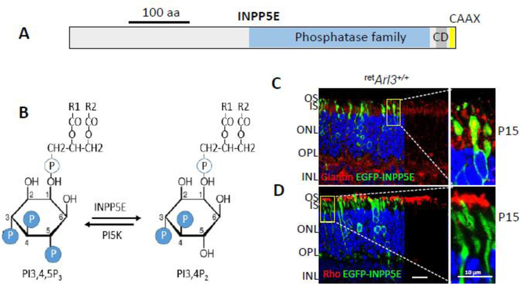
Figure 16.
INPP5E and phosphoinositides. A, INPP5E domain structure. An inositide polyphosphate phosphatase, INPP5E bears a coiled-coil domain at its C-terminal region and a CAAX motif signaling farnesylation. B, INPP5E enzymatically removes a 5’-phosphate at the inositol ring of PI(3,4,5)P3; side chains R1 and R2 are acyl esters attached to glycerol. PI(3,4,5)P3 is a key secondary messenger in photoreceptors and other cells. C, virally-expressed EGFP-INPP5E (green) distributes to the Golgi apparatus of the inner segment and colocalizes partially with the Golgi marker, giantin (red). INPP5E is also found associated with the perinuclear endoplasmic reticulum (ER). D, Anti-rhodopsin labels the rod outer segments where EGFP-INPP5E is undetectable.

