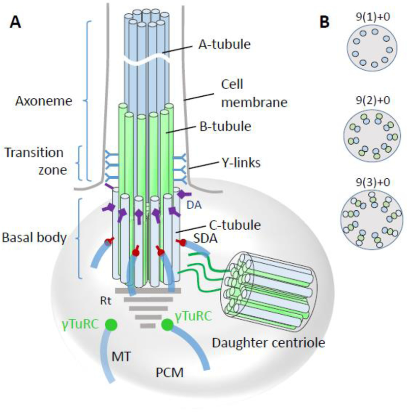
Figure 3.
Basal body/axoneme backbone. A, basal body schematic representation with transition zone and axoneme. A-tubules (blue) and B-tubules (green) emanate from the basal body to form the transition zone which is characterized by y-links connecting the tubules to the ciliary membrane. While the proximal axoneme consists of microtubule doublets, the distal axoneme has singlets. DA, distal appendages; SDA, subdistal appendages; MT, microtubules; PCM, pericentriolar matrix; γT, γ-tubulin ring complex. B, Crossections of the axoneme, transition zone and basal body, distal-to-proximal respectively, indicating the arrangement of microtubule arrays. “+0” indicates absence of a central MT.

